File:PPIIMoL Fig5.jpg
Original file (1,280 × 374 pixels, file size: 62 KB, MIME type: image/jpeg)
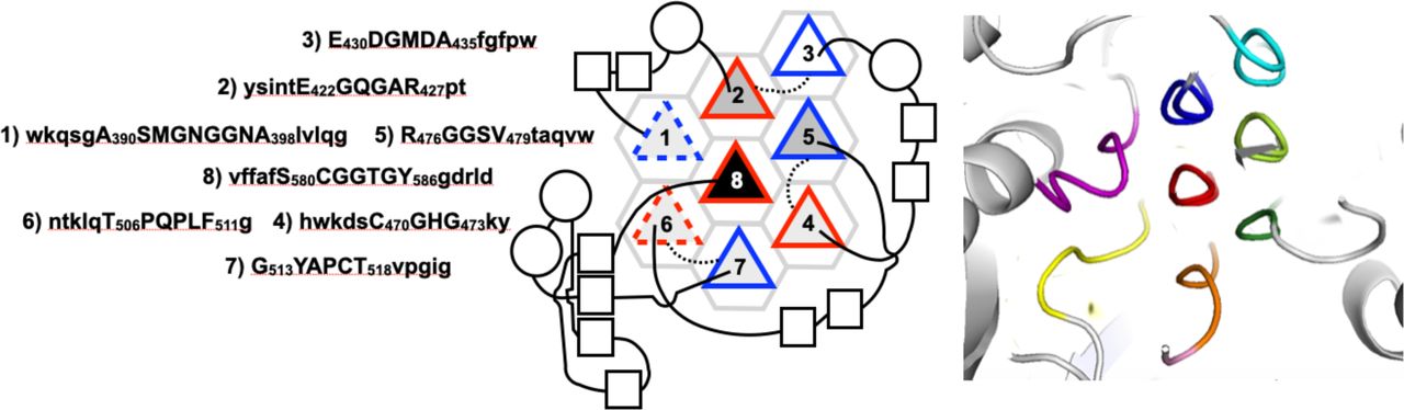
Figure 5. Aromatoleum aromaticum Acetophenone Carboxylase (AaAPC)
A. The primary structure of AaAPC PPII helices is shown, with the PPII helices numbered. Residues forming the PPII helices are written in capital letters; those of flanking sequence are shown in lower case.
B. Schematic diagram representing the PPII helices as triangles. Connecting loops above the plane are shown as solid lines; those below the plane are shown as dotted lines. PPII helical N-termini pointing up are outlined in red; N-termini pointing down are outlined in blue. The dashed outline of PPII helices 1 and 6 refers to their more deviant geometry. PPII helices with two, three, four and six neighboring helices are shaded white, light gray, medium gray and black, respectively. α-helices and β-strands of the connecting segments are represented as circles and squares, respectively. Squares which are aligned and close together represent β-strands hydrogen bonded in β-sheets.
C. The AaAPC tertiary structure (PDB 5L9W) with PPII helices colored purple, blue, cyan, dark green, lime green, yellow, orange and red from the N- to the C-terminus, showing the eight PPII helices end on.
File history
Click on a date/time to view the file as it appeared at that time.
| Date/Time | Thumbnail | Dimensions | User | Comment | |
|---|---|---|---|---|---|
| current | 04:49, 14 August 2025 | 1,280 × 374 (62 KB) | SilviaRodriguez (talk | contribs) |
You cannot overwrite this file.
File usage
The following page uses this file: