File:PPIIMoL Fig4.jpg
Original file (1,280 × 517 pixels, file size: 88 KB, MIME type: image/jpeg)
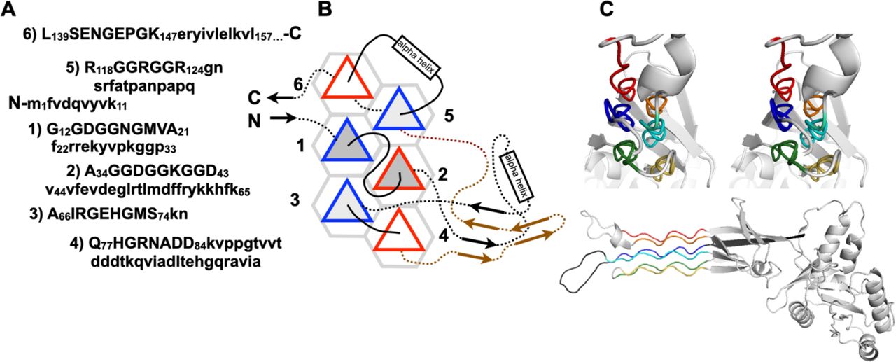
Figure 4. Bacillus subtilis Obg GTP-binding protein (BsObg)
A. Primary structure of BsObg. Residues in PPII helices (which are numbered) are shown in capital letters, others are written in lowercase.
B. Schematic diagram representing the PPII helices as triangles. Connecting loops above the plane are shown as solid lines; those below the plane are shown as dotted lines. For clarity, the connections between PPII helices 4 and 5 are colored brown. PPII helical N- termini pointing up are outlined in red; N-termini pointing down are outlined in blue. PPII helices which two, three and four neighboring helices are shaded white, light gray and medium gray, respectively.
C. Cross-eyed stereo ribbon diagram of the BsObg tertiary structure (PDB 1LNZ) colored blue to red from the N- to the C-terminus, showing the six PPII helices end on (top). The six PPII helices together with the rest of the protein (light gray) are shown in the lower panel.
File history
Click on a date/time to view the file as it appeared at that time.
| Date/Time | Thumbnail | Dimensions | User | Comment | |
|---|---|---|---|---|---|
| current | 04:49, 14 August 2025 | 1,280 × 517 (88 KB) | SilviaRodriguez (talk | contribs) |
You cannot overwrite this file.
File usage
The following page uses this file: