File:PPIIMoL Fig3.jpg
Original file (1,280 × 632 pixels, file size: 118 KB, MIME type: image/jpeg)
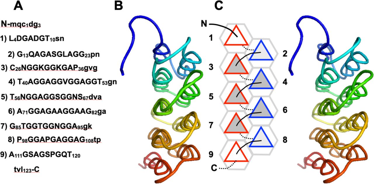
Figure 3. Granisotoma rainieri antifreeze protein
A. Primary structure of GrAFP. Residues in PPII helices (which are numbered) are shown in capital letters, others are written in lowercase.
B. Cross-eyed stereo ribbon diagram of the GrAFP tertiary structure (PDB 7JJV) colored blue to red from the N- to the C-terminus, showing the nine PPII helices end on.
C. Schematic diagram representing the PPII helices as triangles. Connecting loops above the plane are shown as solid lines; those below the plane are shown as dotted lines. PPII helical N-termini pointing up are outlined in red; N-termini pointing down are outlined in blue. PPII helices which two, three and four neighboring helices are shaded white, light gray and medium gray, respectively.
File history
Click on a date/time to view the file as it appeared at that time.
| Date/Time | Thumbnail | Dimensions | User | Comment | |
|---|---|---|---|---|---|
| current | 04:48, 14 August 2025 | | 1,280 × 632 (118 KB) | SilviaRodriguez (talk | contribs) |
You cannot overwrite this file.
File usage
The following page uses this file: