File:PPIIMoL Fig3.jpg: Difference between revisions
No edit summary |
No edit summary |
||
| Line 1: | Line 1: | ||
Figure 3. | |||
Granisotoma rainieri antifreeze protein | |||
A. Primary structure of GrAFP. Residues in PPII helices (which are numbered) are shown in capital letters, others are written in lowercase. | |||
B. Cross-eyed stereo ribbon diagram of the GrAFP tertiary structure (PDB 7JJV) colored blue to red from the N- to the C-terminus, showing the nine PPII helices end on. | |||
C. Schematic diagram representing the PPII helices as triangles. Connecting loops above the plane are shown as solid lines; those below the plane are shown as dotted lines. PPII helical N-termini pointing up are outlined in red; N-termini pointing down are outlined in blue. PPII helices which two, three and four neighboring helices are shaded white, light gray and medium gray, respectively. | |||
Latest revision as of 05:02, 14 August 2025
Figure 3. Granisotoma rainieri antifreeze protein
A. Primary structure of GrAFP. Residues in PPII helices (which are numbered) are shown in capital letters, others are written in lowercase.
B. Cross-eyed stereo ribbon diagram of the GrAFP tertiary structure (PDB 7JJV) colored blue to red from the N- to the C-terminus, showing the nine PPII helices end on.
C. Schematic diagram representing the PPII helices as triangles. Connecting loops above the plane are shown as solid lines; those below the plane are shown as dotted lines. PPII helical N-termini pointing up are outlined in red; N-termini pointing down are outlined in blue. PPII helices which two, three and four neighboring helices are shaded white, light gray and medium gray, respectively.
File history
Click on a date/time to view the file as it appeared at that time.
| Date/Time | Thumbnail | Dimensions | User | Comment | |
|---|---|---|---|---|---|
| current | 04:48, 14 August 2025 | 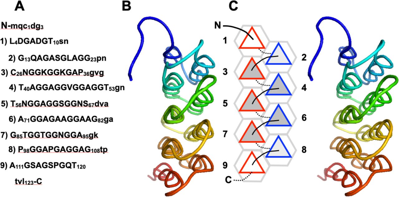 | 1,280 × 632 (118 KB) | SilviaRodriguez (talk | contribs) |
You cannot overwrite this file.
File usage
The following page uses this file: