Difference between revisions of "Cheshift"
| Line 9: | Line 9: | ||
== Description == | == Description == | ||
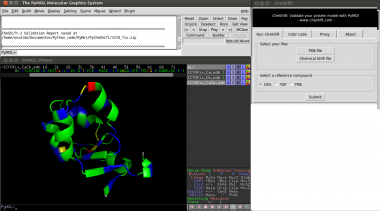
[[Image:cheshift.png|thumb|Result of a CheShift analysis | 380px]] | [[Image:cheshift.png|thumb|Result of a CheShift analysis | 380px]] | ||
| − | The differences between observed and predicted <sup>13</sup>C<sup>α</sup> | + | The differences between observed and predicted <sup>13</sup>C<sup>α</sup> and <sup>13</sup>C<sup>β</sup> chemical shifts can be used as a sensitive probe with which to detect possible local flaws in protein structures. For this reason exist [http://www.cheshift.com CheShift], a Web server for protein structure validation. |
This plugin provides a way to use PyMOL to validate a protein model using observed chemical shifts. All computations run on <i>Che</i>Shift server, | This plugin provides a way to use PyMOL to validate a protein model using observed chemical shifts. All computations run on <i>Che</i>Shift server, | ||
| Line 15: | Line 15: | ||
=== Version === | === Version === | ||
| − | The current version of this plugin is | + | The current version of this plugin is 3.0 |
| Line 44: | Line 44: | ||
3) Select a file with the experimental chemical shift values.<br> | 3) Select a file with the experimental chemical shift values.<br> | ||
4) Click the "Submit" button.<br> | 4) Click the "Submit" button.<br> | ||
| − | 5) Wait until results are displayed (this could | + | 5) Wait until results are displayed (this could from seconds to a few minutes, depending |
| − | on all existing request on the server | + | on all existing request on the server.<br> |
| − | + | ||
<b>Note:</b> | <b>Note:</b> | ||
| − | * If more than 20 conformers are uploaded only the 20 first will be | + | * If more than 20 conformers are uploaded only the 20 first will be analysed. |
| − | * If the PDB file has more than one chain only the first one will be | + | * If the PDB file has more than one chain only the first one will be analysed. |
* The PDB file must not have missing residues. | * The PDB file must not have missing residues. | ||
| − | * Missing observed <sup>13</sup>C<sup>α</sup> chemical shifts are tolerated. | + | * Missing observed <sup>13</sup>C<sup>α</sup> and <sup>13</sup>C<sup>β</sup> chemical shifts are tolerated. |
| − | * | + | * A file with observed <sup>13</sup>C<sup>α</sup> and/or <sup>13</sup>C<sup>β</sup> chemical shift values is needed. The format of this file should be the one used in the BMRB or the PDB. Alternatively, you can provide a simple 4 column file : The first column should be the residue number, the second the residue name (three-letter code) and the third column the <sup>13</sup>C<sup>α</sup> experimental chemical shifts and the last column the <sup>13</sup>C<sup>β</sup> chemical shifts. Spaces should be used to separates the columns. |
| − | |||
| − | |||
| − | |||
| − | |||
| − | |||
| − | |||
| − | |||
| + | 1 MET 55.63 32.95 | ||
| + | 2 TYR 62.81 39.27 | ||
| + | 3 ALA 53.78 18.97 | ||
| + | 4 GLY 47.24 999.00 | ||
| + | 5 LYS 57.55 32.77 | ||
| + | 6 ILE 56.38 38.59 | ||
=== Proxy Configuration === | === Proxy Configuration === | ||
| Line 73: | Line 72: | ||
== Citation == | == Citation == | ||
| − | Martin O.A. | + | Martin O.A. Arnautova Y.A. Icazatti A.A. Scheraga H.A. and Vila J.A. A Physics-Based Method to Validate and Repair Flaws in Protein Structures. Proc Natl Acad Sci USA 2013. <i>(in press). |
| − | |||
| − | + | Martin O.A. Vila J.A. and Scheraga H.A. (2012). <i>Che</i>Shift-2: Graphic validation of protein structures. Bioinformatics 2012. 28(11), 1538-1539. | |
| − | Martin O.A. Vila J.A. and Scheraga H.A. (2012). <i>Che</i>Shift-2: Graphic validation of protein structures. Bioinformatics 2012. | ||
| + | == Other References == | ||
Vila J.A. Arnautova Y.A. Martin O.A. and Scheraga, H.A. (2009). Quantum-mechanics-derived 13C chemical shift server (<i>Che</i>Shift) for protein structure validation. PNAS, 106(40), 16972-16977. | Vila J.A. Arnautova Y.A. Martin O.A. and Scheraga, H.A. (2009). Quantum-mechanics-derived 13C chemical shift server (<i>Che</i>Shift) for protein structure validation. PNAS, 106(40), 16972-16977. | ||
Revision as of 18:18, 28 August 2013
| Type | PyMOL Plugin |
|---|---|
| Download | plugins/cheshift.py |
| Author(s) | Osvaldo Martin |
| License | GPL |
| This code has been put under version control in the project Pymol-script-repo | |
Description
The differences between observed and predicted 13Cα and 13Cβ chemical shifts can be used as a sensitive probe with which to detect possible local flaws in protein structures. For this reason exist CheShift, a Web server for protein structure validation.
This plugin provides a way to use PyMOL to validate a protein model using observed chemical shifts. All computations run on CheShift server, and the results are retrieved to PyMOL, hence an Internet connection is needed.
Version
The current version of this plugin is 3.0
Installation
Linux
1) The plugin can be downloaded from here [Pymol-script-repo]
2) You should install mechanize, a python module. This module is available from the repositories of the main Linux distributions. Just use your default package manager (or command line) to install it. In Ubuntu/Debian the package name is "python-mechanize".
Windows
This plugin have not been extensively tested on Windows machines, but it passed all the test I have done...
1) The plugin can be downloaded from here [Pymol-script-repo]
2) You should install mechanize, a python module. In order to do that, download this unzip and copy the mechanize folder were you have installed PyMOL, usually is C:\Program Files\DeLano Scientific\PyMOL\modules\pmg_tk\startup
Mac OsX
This plugin have not been tested on a Mac OsX machine, but it should work...
1) The plugin can be downloaded from here [Pymol-script-repo]
2) You should install mechanize, a python module. In order to do that download this unzip and copy the mechanize folder were you have installed PyMOL. For the X11/Hybrid version, the location is probably: PyMOLX11Hybrid.app/pymol/modules/pmg_tk/startup
Using the Plugin
1) Launch PyMOL and select "Cheshift" from the plugin menu.
2) Select a PDB file of your protein model (using the CheShift plugin menu).
3) Select a file with the experimental chemical shift values.
4) Click the "Submit" button.
5) Wait until results are displayed (this could from seconds to a few minutes, depending
on all existing request on the server.
Note:
- If more than 20 conformers are uploaded only the 20 first will be analysed.
- If the PDB file has more than one chain only the first one will be analysed.
- The PDB file must not have missing residues.
- Missing observed 13Cα and 13Cβ chemical shifts are tolerated.
- A file with observed 13Cα and/or 13Cβ chemical shift values is needed. The format of this file should be the one used in the BMRB or the PDB. Alternatively, you can provide a simple 4 column file : The first column should be the residue number, the second the residue name (three-letter code) and the third column the 13Cα experimental chemical shifts and the last column the 13Cβ chemical shifts. Spaces should be used to separates the columns.
1 MET 55.63 32.95 2 TYR 62.81 39.27 3 ALA 53.78 18.97 4 GLY 47.24 999.00 5 LYS 57.55 32.77 6 ILE 56.38 38.59
Proxy Configuration
If you are behind a proxy the CheShift plugin will try to correctly guess your proxy configuration, but this is a tricky business and many things could fail. In that scenario you will be prompted to manually set your proxy settings. In case you manually set the proxy configuration the plugin will save your proxy settings and next time it will attempt to use those saved settings to connect to the Internet.
License
CheShift plugin is free software: you can redistribute it and/or modify it under the terms of the GNU General Public License. A complete copy of the GNU General Public License can be accessed here http://www.gnu.org/licenses/.
CheShift Server (www.cheshift.com) is available free of charge ONLY for academic use.
Citation
Martin O.A. Arnautova Y.A. Icazatti A.A. Scheraga H.A. and Vila J.A. A Physics-Based Method to Validate and Repair Flaws in Protein Structures. Proc Natl Acad Sci USA 2013. (in press).
Martin O.A. Vila J.A. and Scheraga H.A. (2012). CheShift-2: Graphic validation of protein structures. Bioinformatics 2012. 28(11), 1538-1539.
Other References
Vila J.A. Arnautova Y.A. Martin O.A. and Scheraga, H.A. (2009). Quantum-mechanics-derived 13C chemical shift server (CheShift) for protein structure validation. PNAS, 106(40), 16972-16977.
Vila, J.A. and Scheraga H.A. (2009). Assessing the accuracy of protein structures by quantum mechanical computations of 13Cα chemical shifts. Accounts of chemical research, 42(10), 1545-53.
